High tech camera provides researchers with a potentially powerful screening tool for premature babies
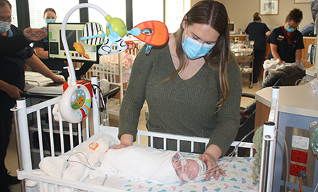
 Charlotte Buxton pictured with her baby son Angus who participated in the Emubird study
Researchers in the Child and Adolescent Health Service’s Neonatal Unit at King Edward Memorial Hospital’s (KEMH) are leading an Australian first study to use powerful images of a baby’s eye as a window into their developing brain.
The ‘Emubird’ study led by Neonatologist Dr Rolland Kohan has been made possible thanks to Telethon Trust funding for a handheld retinal optical device or camera that gives researchers a 3-dimensional view of the retina behind their eye.
Dr Kohan said the aim of the study was to identify images of a baby’s retina which could predict developmental concerns and therefore alert clinicians to a need for closer screening as the baby grows.
“Early detection and early intervention are key to good outcomes in children so it would be exciting for us to contribute to the existing suite of screening tools to help give these babies the best possible start to life,” he said.
The study has recently achieved a significant halfway milestone with 50 babies recruited. All babies on the study must be under 29 weeks gestational age at birth.
“This camera, which has been used to screen adults for macular degeneration for more than a decade, is incredibly powerful, because the images it provides of the retina are 10 times clearer than images from a Magnetic Resonance Imaging scan,” said Dr Kohan.
“We are the first neonatal unit in Australia to use the device at the cot side which gives us a view of the developing retina never previously available.
“This camera provides additional information beyond the existing RetCam imaging used to routinely screen all babies for Retinopathy of Prematurity (ROP).”
Charlotte Buxton’s son Angus is one of the babies who has participated in the study. Angus has been cared by staff in the neonatal intensive care unit at KEMH since his birth at 26 weeks.
“I am so happy to support this research because I’m mindful we have benefited enormously from research studies in the past and families who have gone before,” Mrs Buxton said.
The retina, which is the innermost layer of the eye, and the brain normally develop inside the womb so premature babies therefore risk developing ROP and developmental delays.
Babies participating in the study will continue to be monitored for their first two years of life in order to compare their developmental data with the information detected from their eye scans as a newborn.
“We are grateful to all of the parents who have consented to be part of this study for their important contribution to helping inform medical evidence which will benefit patients and families for years to come,” Dr Kohan said.
The study is funded by Telethon Trust and is being conducted in collaboration with Perth Children’s Hospital colleagues Clinical Associate Professor Geoffrey Lam and Professor Lakshmi Nagarajan, Co-Director Neonatology Associate Professor Mary Sharp and University of Western Australia Associate Professor Barry Cense.
Find out more about this project by watching this story from the Telethon broadcast.

